Silent sinus syndrome (SSS) is a rare condition that can pose a diagnostic challenge. The patient may present with unilateral
ptosis or retraction, a deep superior sulcus or orbital asymmetry.The medical history is often non-contributory. This condition
is characterized by unilateral spontaneous enophthalmos and hypoglobus due to increased orbital volume and retraction of the orbital floor. This occurs because of atelectasis of the ipsilateral maxillary sinus and when the condition is left untreated, may result in complete obliteration of the sinus with worsening enophthalmos and hypoglobus.The first two cases were reported in 1964, but the term “SSS” was coined 30 years later by Soparkar et al. Since that time, several case series have been published in both the ophthalmology and otolaryngology literature. To our knowledge, a number of these case reports in radiology literature are less and many radiologists remain unfamiliar with the syndrome and its characteristic radiological findings. In this article, we discuss the presentation, diagnosis, pathogenesis, and treatment of SSS.MRI of the patient was carried out for further evaluation and to rule out underlying mass lesion. The T1- and T2-weighted images [Figures 2-4] showed downward displacement of the right orbital floor with resulting increase in orbital volume. Right maxillary sinus was seen to be reduced in volume. However, it was completely opacified. There was also noted inward retraction of posterior and medial walls of maxillary sinus which was causing reduction in its volume [Figure 5]. So in view of clinical and imaging findings, diagnosis of SSS was given. Computed tomography (CT) was not performed as MRI was sufficient as a diagnostic tool. Patient did not have any other complaints other than orbital asymmetry, so he refused to undergo functional endoscopic sinus surgery (FESS) or other therapeutic procedures.
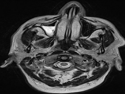
Figure 2: Axial T2-weighted images MRI image showing fluid level in
right maxillary sinus with reduction in its volume
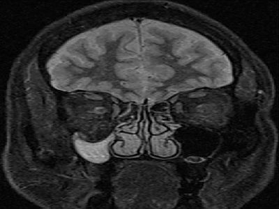
Figure 4: Coronal fluid attennuated inversion recovery (FLAIR) image
showing reduction in volume of right maxillary sinus with inward
retraction of its walls
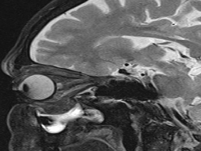
Figure 3: Sagittal T2-weighted images MRI image shows reduction
in volume of right maxillary sinus with inward retraction of its walls
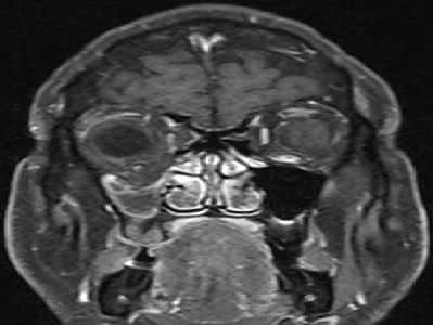
Figure 5: T1-weighted fat-saturated post-contrast MR image obtained
in coronal plane displays inward retraction of posterior and medial walls
of right maxillary sinus. There is also downward retraction of orbital
floor into sinus lumen